
Coordinator: Piernicola Oliva
Phone: + 39 079 / 229485 – 320/4329030
Email: oliva@uniss.it
PHYSICS APPLIED TO MEDICINE
Bruno Golosio – golosio@unica.it
Viviana Fanti – viviana.fanti@ca.infn.it
Piernicola Oliva – oliva@uniss.it
Technological and applied research is coordinated at the Italian level by the National Scientific Commission 5.
Diagnostic imaging
The Cagliari Unit has always been involved in the use of X-rays for medical diagnostics. In conventional radiology, the group has expertise in the characterization of X-ray sources and detectors, both for planar imaging (in projection) and in three-dimensional imaging (computerized axial tomography, TAC).
The Division of Cagliari is also dedicated to the development of reconstruction and processing techniques of biomedical images.
In addition to conventional imaging, in which the absorption of x-rays by the material is exploited, the group has been working for years on phase contrast imaging, in which the undulatory properties of X-rays and the different phase shift of these are exploited. waves from different materials, to produce images. In addition to the X-ray tubes, the group operates on high-brightness sources, such as synchrotrons or reverse Compton sources, working on the characterization of these sources and their possible medical applications.
The Cagliari Unit works in the analysis of magnetic resonance images (MRI), looking for biomarkers for neurodevelopmental or neurodegenerative pathologies. MRI is based on the physical process of nuclear magnetic resonance; it also allows to obtain three-dimensional images and, compared to the TAC, it is able to better discriminate the soft tissues, and has the advantage of not using ionizing radiations.
PET is a technique based on the patient’s administration of radioactive substances linked to appropriate biological molecules, which provides important functional information. The most widely used radiopharmaceutical, for example, is based on a sugar that accumulates in tumors and makes it possible to diagnose lesions at a very early stage. The Cagliari group is involved in projects on innovative PET detectors.
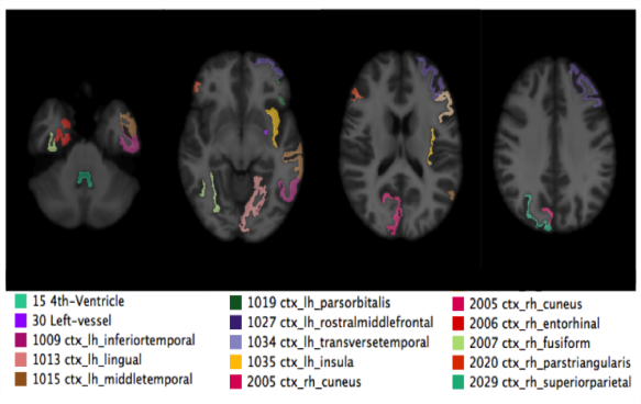
In the figure: an example of a map of the most discriminating regions between patients with autism spectrum disorder and controls in magnetic resonance, evaluated using machine learning techniques.
Simulation of the radiation-matter interaction and dosimetry
Many biomedical imaging techniques are based on processes of interaction of radiation with matter and / or processes of decay of radioactive nuclei. Knowing these processes is essential for the design of imaging systems, for the development of reconstruction and image analysis software and for patient dose estimates.
The Cagliari Unit deals with the development and application of simulation software for radiation interaction processes with matter and decay processes, using the Monte Carlo method, based on random sampling of the possible results of each event.
Radiotherapy
Radiotherapy consists of the use of ionizing radiation for the treatment of diseased tissues, especially tumors. To reduce the dose delivered to healthy tissues and to maximize the amount deposited in tumors, advanced techniques have been developed, based on the use of different types of radiation. Cagliari participates in innovative radiotherapy projects, in collaboration with international healthcare facilities and research centers.
Neuroscience
The field of neuroscience research is an interdisciplinary field, to which physicists also contribute thanks to their expertise on artificial neural networks and neural signal models on the one hand, and on experimental techniques (for example, functional magnetic resonance imaging and electrophysiological recordings). ) on the other.
One of the great challenges of research in the coming decades, on which the Cagliari group is also involved, will be to try to understand how high-level cognitive functions, such as those associated with language and reasoning, are linked to the transmission of signals between the neurons of our brain. This research activity is important not only because it helps us to understand how our brain works, but also because it can help us in the study of neurological disorders and in the research of diagnosis and therapy techniques of these disorders.
PHYSICS APPLIED TO CULTURAL HERITAGE
Piernicola Oliva – oliva@uniss.it
On cultural heritage, the Cagliari Unit has been using elemental characterization techniques such as XRF (X-Ray Fluorescence) for many years, in which the characteristic emissions of ionized atoms are exploited to identify the elements present in a sample and their concentrations.
However, the XRF has some limitations due to the low energy of the X-rays emitted. For some elements this energy is so low that the X-rays are completely absorbed (from the material itself, from the air or from the detector window) before they can be detected. For other elements, even if the X-rays emitted have sufficient energy to be detected, their attenuation makes it difficult to obtain reliable quantitative information. This is why XRF is considered a surface investigation technique.
A possible alternative for elemental characterization is muon spectroscopy. In this technique atomic electrons are replaced by negative muons. The photons emitted by the de-excitation of these atoms have energies ~ 200 times greater than those of fluorescence X, do not undergo self-absorption and are perfectly detectable, allowing elemental characterization even in depth and also for low concentrations. Measurements are performed at the Rutherford Appleton Laboratory (UK).
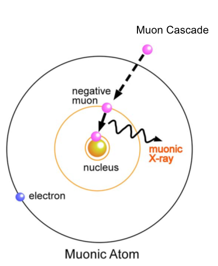
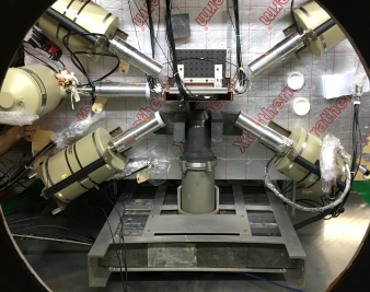
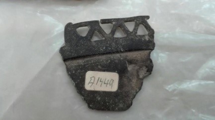
In the picture: a muon atom, the experimental setup at the RAL and a bronze nuragic family.
TIMESPOT
National Responsible: Adriano Lai, INFN Cagliari, adriano.lai@ca.infn.it
Website: https://web.infn.it/timespot/
Collider experiments are preparing to face the challenge of high brightness, which poses new technological requirements, especially with regard to vertex detection and tracking techniques. These consist of the high spatial resolution, already typical of vertex detectors (50-100 μm), but now it is coupled to a resistance to radiation fluxes of some 1016 1 MeV equivalent neutrons per cm2 and to the possibility of a measurement of the high resolution time (<50 ps) at the level of the single pixel. This last requirement is necessary to combat the high pile-up generated by the high brightness which poses great complications to the exact identification of the vertices and reconstruction of the events. In addition, the high flow of data exiting the detector (in the order of Tbits / s) requires an early reduction of data and possibly the use of real-time reconstruction techniques.
The TIMESPOT project (acronym for TIME and SPace real-time OPerating Tracker) was funded by the CSN5 for the years 2018-2020 with a budget of around 1 million euros. The final objective of the project is to create a demonstration tracer apparatus (telescope with at least 4-5 tracking planes) that integrates 3D sensors (silicon and diamond) with high spatial and temporal resolution, integrated electronics for reading sensor matrices and time measurement for every pixel in 28 nm CMOS technology, calculation systems that realize real-time reconstruction algorithms.
The project brings together 10 INFN Sections (Bologna, Cagliari, Ferrara, Florence, Genoa, Milan, Padua, Perugia, Turin, Trento) and many of the leading INFN experts in the various sectors of activity involved. It tries to go beyond the current limit of already practicable tracking techniques, to prepare the new generation of experiments to high-brightness colliders.
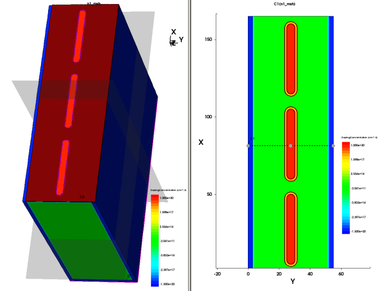
Figure: 3D high resolution temporal sensor model currently in production